
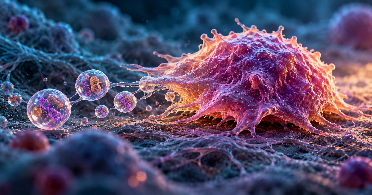
A cancer cell is moving across a surface. Behind it, a thin strand of its membrane stretches like a thread. Then a tiny bubble breaks free. Inside that bubble are pieces of cellular machinery—and DNA carrying the genetic signature of the cell that released it.
This is not a scene from science fiction. In research published on October 28, 2026, scientists described a previously uncharacterized class of extracellular vesicles produced during cancer cell migration. They called them motile vesicles, or MoVes. The discovery reveals an unexpected link between the way certain cancer cells move and the material they leave behind. Nature Cell Biology: Original study
But there is an important limit to the finding: discovering a mechanism that releases cancer-related DNA is not the same as proving that it causes cancer to spread in patients.
Cells can send packages without moving themselves
Our cells are constantly exchanging information. One way they do this is by releasing extracellular vesicles: small membrane-bound packages that can carry proteins, RNA, DNA, and other components. Depending on where they come from and what they contain, these packages may influence the behavior of cells that encounter them. Research Institute of the McGill University Health Centre: Discovery announcement
Think of a cell as a busy workshop. It does not have to send the entire building to deliver a message; sometimes it sends a sealed parcel instead. That analogy is useful, but imperfect. These biological parcels are not necessarily instructions, and scientists must test what they actually do after release.
Cancer researchers have long studied extracellular vesicles because some can influence the environment around tumors. The new finding adds a distinct possibility: a cell’s movement itself can help create an unusual type of package.
The surprise was at the back of the moving cell
The research team, led by Julia V. Burnier at the Research Institute of the McGill University Health Centre, was investigating how cancer cells release DNA. Using live-cell imaging and electron microscopy, the researchers saw large vesicles forming as migrating cells pulled their rear edges forward.
During migration, the trailing edge can stretch into thin connections. In the observations reported by the researchers, vesicle-like structures developed along these extensions and then detached. The MoVes measured roughly 2–5 micrometers across—large compared with many extracellular vesicles. Nature Cell Biology: Study and microscopy figures
That sequence matters. The researchers were not merely finding loose DNA near a tumor cell. They were watching a defined structure emerge during cell movement, become separated from its parent cell, and carry biological material away.
The team compared cancerous and non-transformed cell models. MoVes were more readily observed in the migrating cancer-cell systems studied and were rare in the non-transformed cells under the reported conditions. This is a laboratory observation, not a claim that healthy cells can never produce similar structures.
What is inside a MoVe?
This is where the story gets especially striking.
Genomic analysis found that MoVes carry DNA spanning the parent cell’s genome and reflecting its characteristic mutations. They also contained cellular organelles and proteins. Some of the structures identified included mitochondria, the compartments involved in producing much of a cell’s usable energy. Nature Cell Biology: Original study
It is tempting to call them miniature cancer cells. That would be misleading. A MoVe is a released membrane-bound structure, not a new independently reproducing tumor cell. Finding DNA across the genome also does not establish that every single vesicle contains a pristine, complete copy of every chromosome.
The researchers investigated what the cargo might do. In experiments reported in the paper, MoVe-associated mitochondrial material could increase oxidative phosphorylation—a cellular energy-producing process—in recipient cells. That is a measurable biological effect, but it is not equivalent to demonstrating that a vesicle creates a metastatic tumor. Nature Cell Biology: Functional findings
Why movement changes the picture
Some cells shift into a more mobile state through a process often called epithelial-to-mesenchymal transition (EMT). In simplified terms, cells that normally form cohesive layers can acquire characteristics that help them move through tissue. EMT is not exclusive to cancer; it has roles in normal biology, too.
The researchers found that MoVe production was associated with this transition. When they induced more mobile behavior in a non-cancerous cell model, MoVe production increased. That result supports the idea that the underlying cell state and migration mechanics help generate these vesicles. McGill University Health Centre: Researcher explanation
It does not mean that EMT always produces MoVes, that MoVes are unique to malignant cells, or that measuring them would currently tell a doctor how an individual cancer will progress.
Does this explain how cancer spreads?
Not yet. The answer depends on what we mean by spreads.
Metastasis occurs when cancer cells leave their original site, travel to another part of the body, and establish new tumors. It is a complicated, multistep process, not merely the movement of cancer-related material. National Cancer Institute: Metastatic Cancer
MoVes may become relevant to that process because they can transport genetic material and organelles. The researchers also explored how MoVes behave experimentally in animal models. Yet the published work does not establish that naturally released MoVes initiate metastases in patients.
A useful way to separate the evidence is this:
| What the study supports | What remains unknown |
|---|---|
| Migrating cancer cells can shed a distinct type of large vesicle | How common MoVes are across human cancers |
| MoVes carry genomic DNA and other cellular cargo | Whether MoVes directly drive metastasis in patients |
| Some MoVe cargo affects recipient-cell energy metabolism in experiments | Whether MoVes will be useful for diagnosis or treatment |
This distinction is what makes early-stage research worth understanding rather than overselling.
Could doctors one day use these particles?
Possibly—but that is a research question, not a clinical promise.
The McGill team says it wants to learn more about the mechanisms governing MoVe formation, the effects of their cargo, and whether these structures can be detected in animal models and patients. If a reliable relationship to disease progression eventually emerges, they might be investigated as biomarkers: measurable clues about a biological process. Research Institute of the McGill University Health Centre: Next research steps
A biomarker, however, needs more than an interesting laboratory signal. Researchers would have to show it can be measured reliably, distinguishes clinically meaningful situations, and adds useful information beyond existing tests. None of that has been established for MoVes.
There is also no new treatment for patients to request on the basis of this discovery. Current cancer care should not change because of an unvalidated experimental finding.
The bigger lesson: cells leave more than footprints
What makes this discovery fascinating is not an immediate medical breakthrough. It is the picture of a moving cell physically shedding packets of information and machinery as part of its journey.
For decades, researchers have studied how cells migrate and how they communicate. MoVes offer a new place where those two fields meet: the mechanics of movement may shape what a cell releases to its surroundings.
The next question is the decisive one. Do these packages meaningfully change what other cells do inside a living tumor—and, if so, how? Until that is answered, the discovery is best described as a new biological mechanism with promising questions attached, not a solved mystery of metastasis.
Sometimes understanding a disease begins with noticing something tiny that was hiding in plain sight: not the cell moving forward, but what it leaves behind.
Sources
- Tsering, T. et al. (2026) — Motile vesicles (MoVes) are a cancer cell migration-associated extracellular vesicle population. Nature Cell Biology.
- Research Institute of the McGill University Health Centre — Discovery of a potential new vehicle for cancer spread (October 28, 2026).
- McGill University Health Centre — Research announcement (October 28, 2026).
- National Cancer Institute — Metastatic Cancer: When Cancer Spreads.